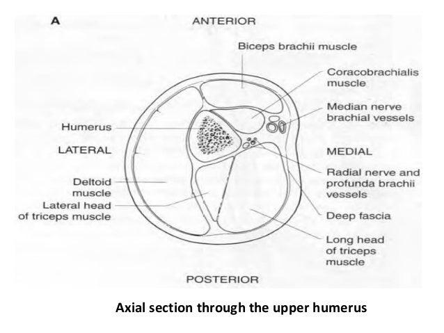
Hand Muscles Mri Anatomy
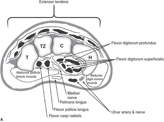
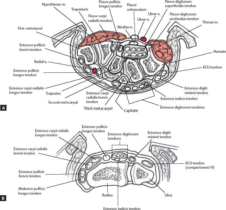
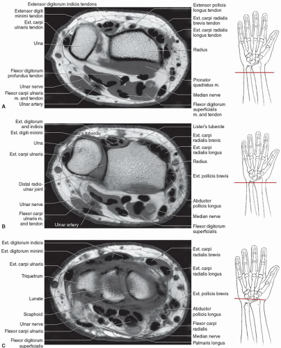
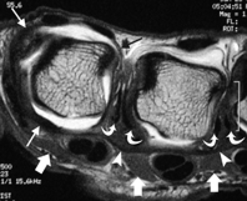
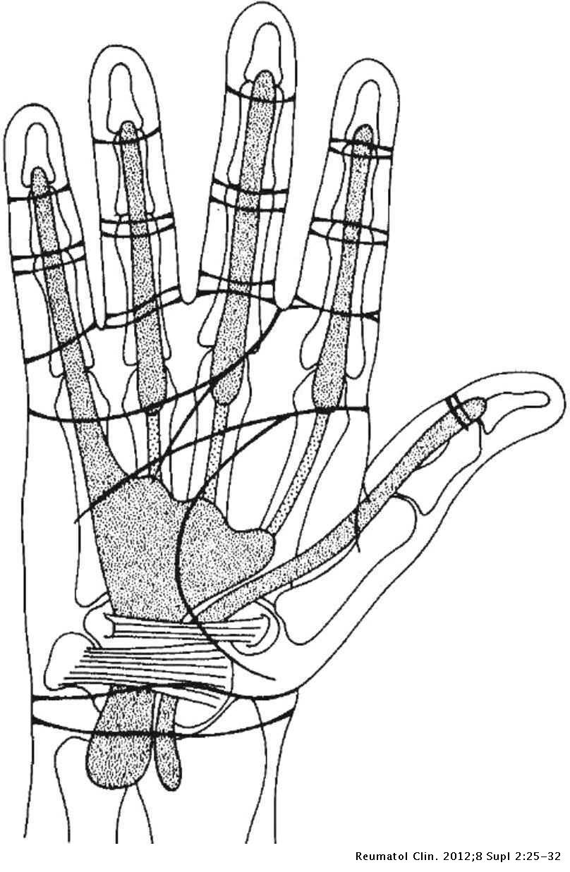
Anatomy of the hand and the wrist on mri cross sectional anatomy of the hand on mr imaging wrist metacarpus fingers the hand mr.

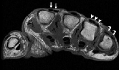
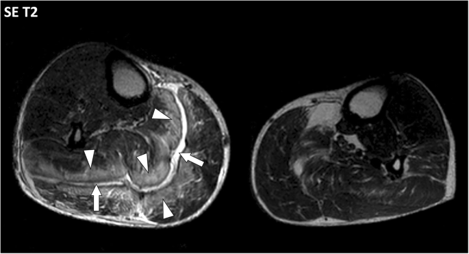
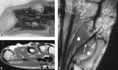
Hand muscles mri anatomy. Although representing incidental normal variants anomalous intrinsic and extrinsic hand muscles are pitfalls on mri. Variation in the muscles bellies and attachments of the lumbricals are common with any being possibly unipennate or bipennate. Mri of 21 hands was performed in 19 patients with clinically evident or suspected intrinsic hand muscle abnormalities. The intrinsic muscles of the hand are located within the hand itself.
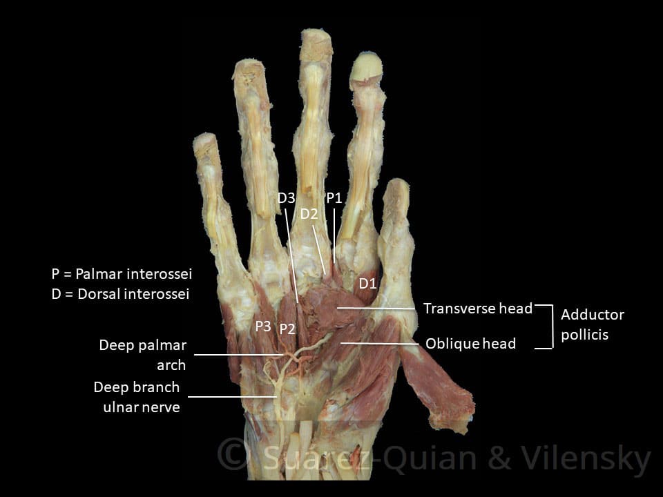
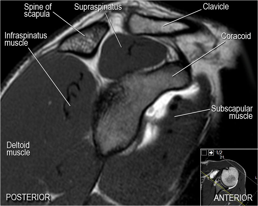
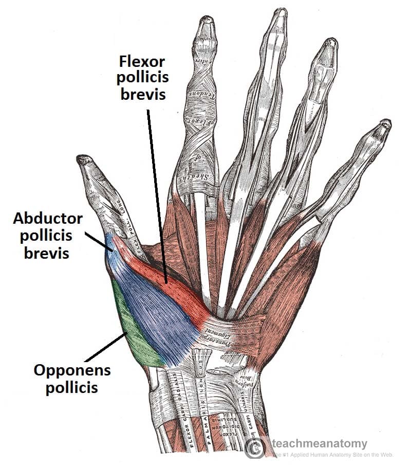
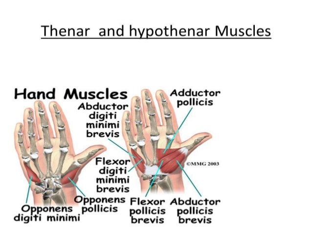
Medical imaging anatomy atlas. Mri of the upper extremity anatomy atlas of the human body using cross sectional imaging we created an anatomical atlas of the upper limb an interactive tool for studying the conventional anatomy of the shoulder arm forearm wrist and hand based on an axial magnetic resonance of the entire upper limb. This webpage presents the anatomical structures found on wrist mri. In this article we shall be looking at the anatomy of the intrinsic muscles of the hand.
Atlas of wrist mri anatomy. These include the adductor pollicis palmaris brevis interossei lumbricals thenar and hypothenar muscles. They are responsible for the fine motor functions of the hand. Muscles of the hand anatomy tutorial duration.
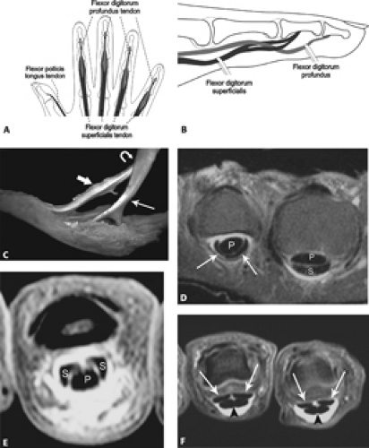
Click on a link to get t1 axial view t1 coronal view. Use the mouse to. All mri was performed on a 15 t scanner using transaxial t1 weighted t2 weighted or stir as well as contrast enhanced t1 weighted sequences. The lumbrical muscles of the hand are intrinsic muscles of the hand associated with the flexor digitorum profundus fdp tendon.
Intrinsic muscles of the hand explained. The hand itself consists of specific bones onto which various muscles are attached and a.
:background_color(FFFFFF):format(jpeg)/images/library/12304/mri-pd-axial-glenoid-cavity-level_english.jpg)






:background_color(FFFFFF):format(jpeg)/images/library/8869/mri-t1-axial-tendon-extensor-carpi-ulnaris-level_english.jpg)


:background_color(FFFFFF):format(jpeg)/images/library/12298/mri-t2-axial-caudate-nucleus-level_english.jpg)










:background_color(FFFFFF):format(jpeg)/images/library/12305/mri-t1-axial-carpal-tunnel_english.jpg)





:background_color(FFFFFF):format(jpeg)/images/library/10610/DorsalHand_Thumbnail.png)


