Hand Anatomy Tendons Dorsal
The dorsal wrist ligaments are comparatively thin.
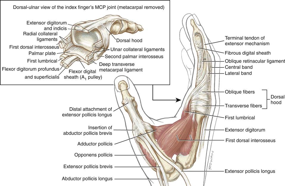
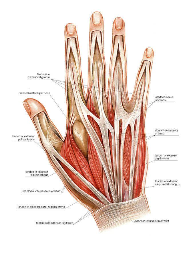
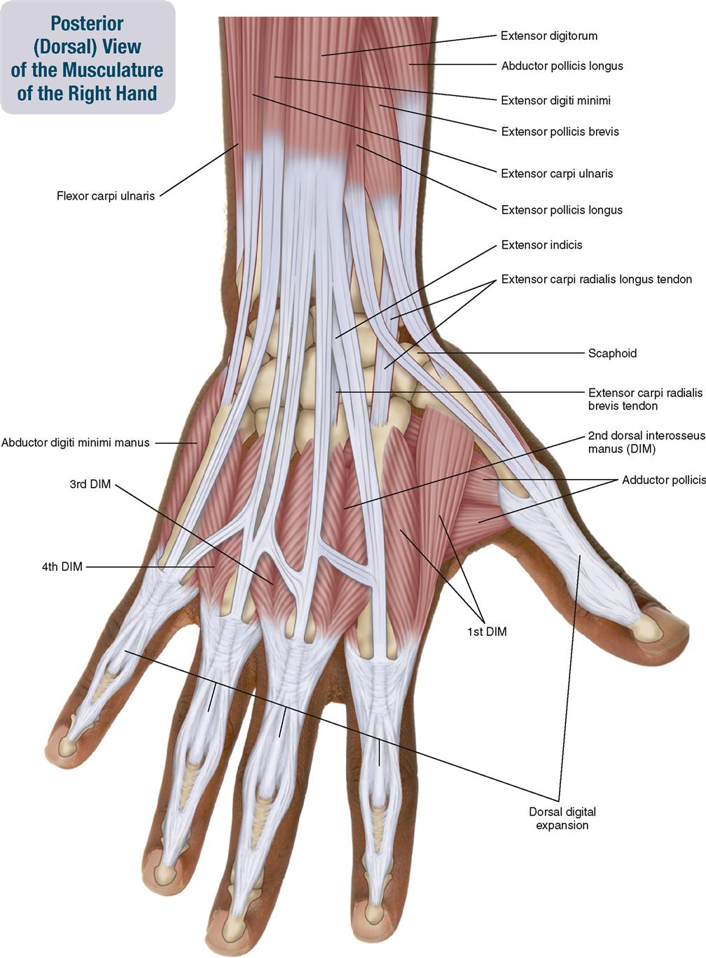
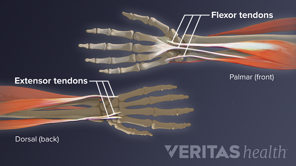
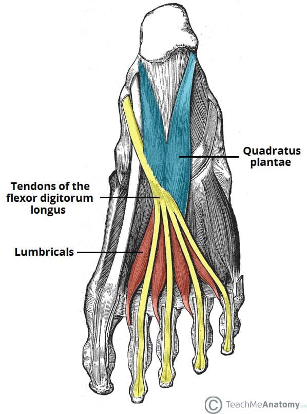
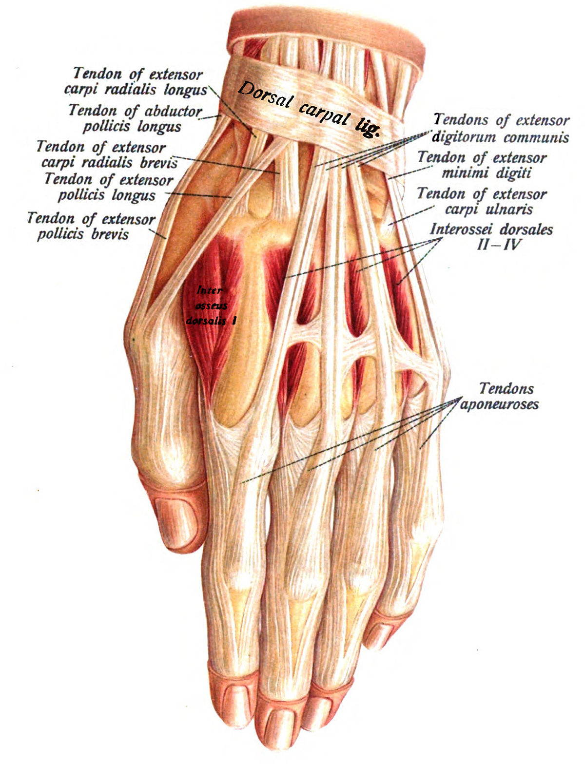
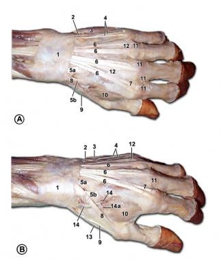
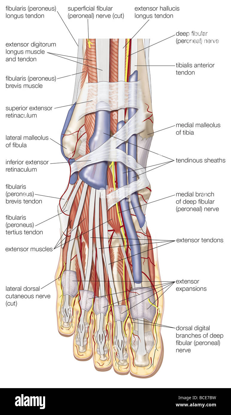
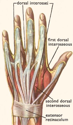
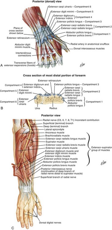
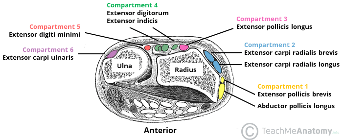
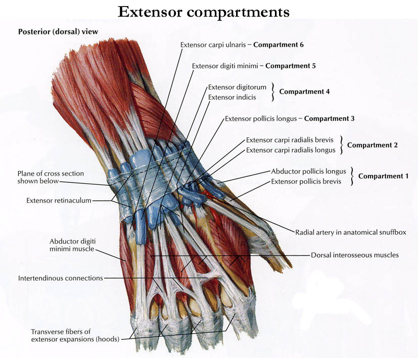
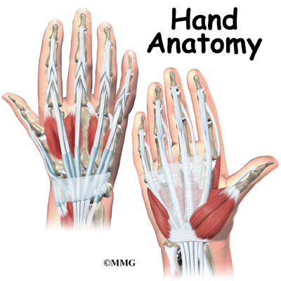
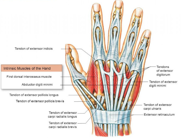
Hand anatomy tendons dorsal. A hand is a prehensile multi fingered appendage located at the end of the forearm or forelimb of primates such as humans chimpanzees monkeys and lemursa few other vertebrates such as the koala which has two opposable thumbs on each hand and fingerprints extremely similar to human fingerprints are often described as having hands instead of paws on their front limbs. Toes are digits too but notice the absence of longus or brevis here. The tendons that allow each finger joint to straighten are called the extensor tendons. The tendons of the dorsal wrist are also separated into six fibro osseous compartments see fig.
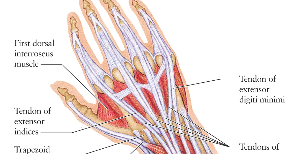
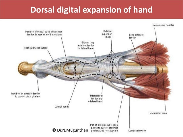
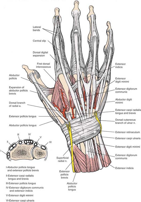
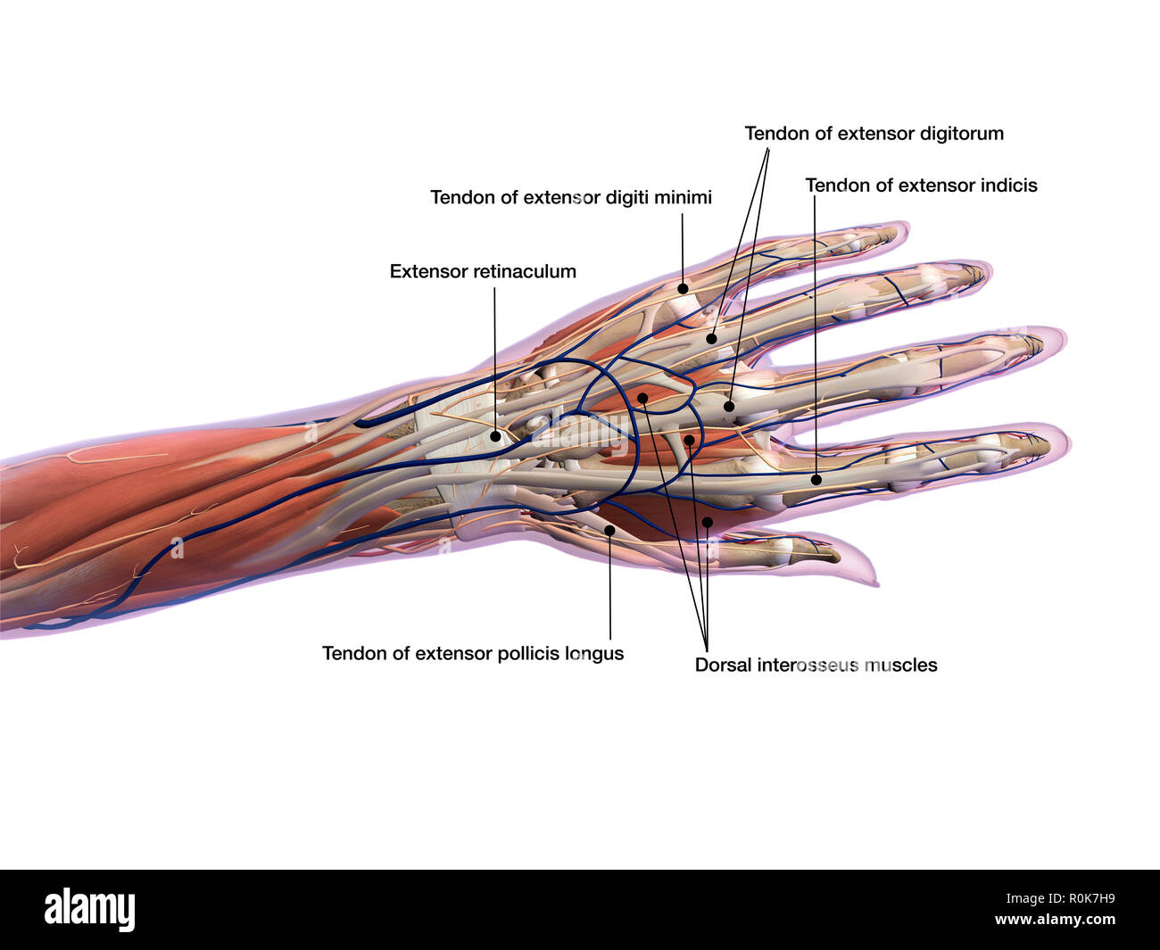
The four tendons then continue along the back of the hand and onto each finger. Reinforced by the floor and septa of the fibrous tunnels for the six dorsal compartments see below and have a z shaped configuration. The easiest tendons to identify in the dorsal hand are those of the extensor digitorum muscle. For more anatomy content please follow us and visit our website.
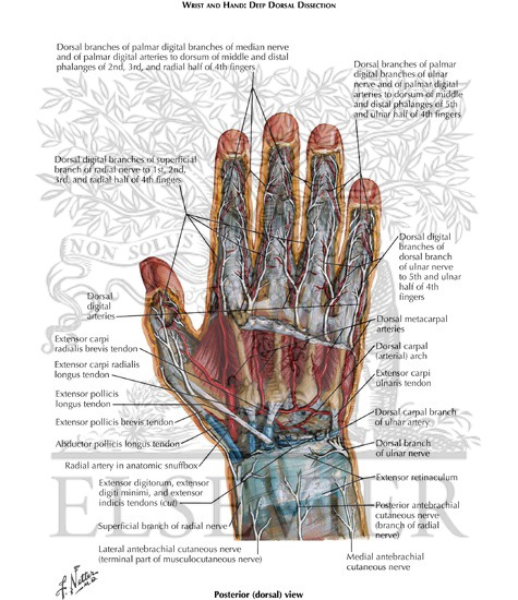
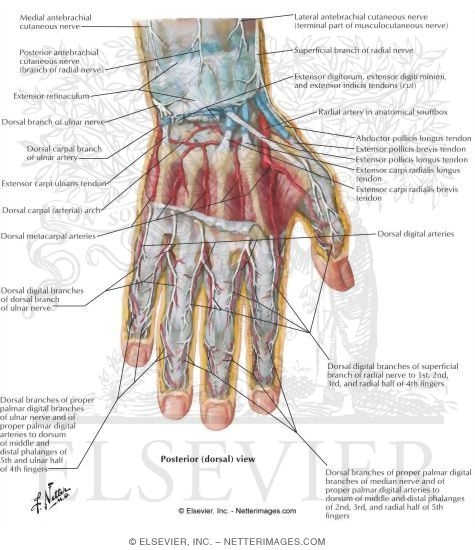
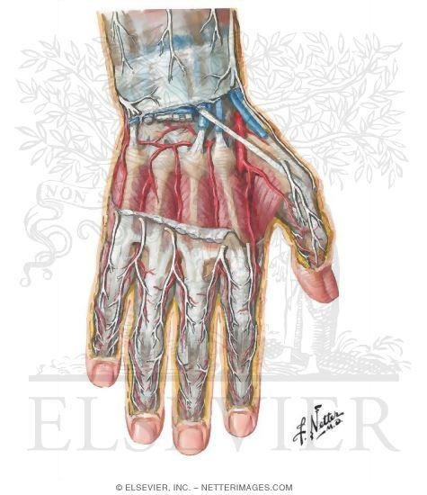
The fibres of the dorsal radiocarpal ligaments are aligned more or less in the same axis as the forearm those of. Unlike the toes the fingers have only one extensor muscle so it needs no further qualifier in its name. Extensor digiti minimi edm tendon. We are pleased to provide you with the picture named dorsal hand tendon nerve artery vein anatomywe hope this picture dorsal hand tendon nerve artery vein anatomy can help you study and research.
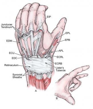
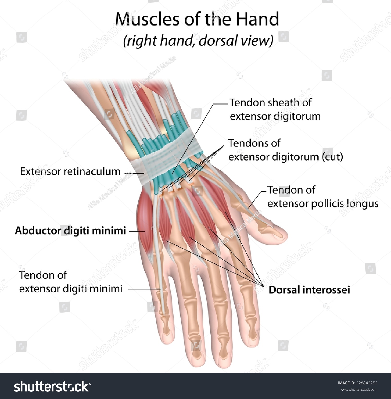
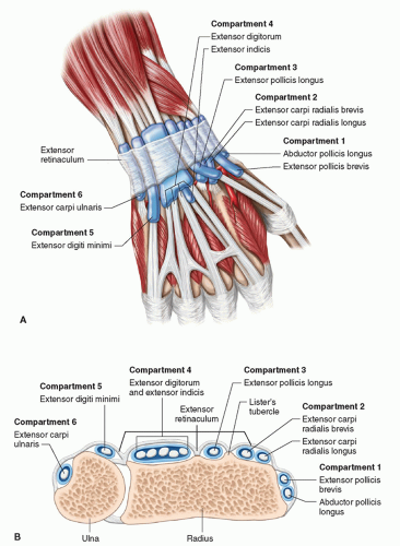
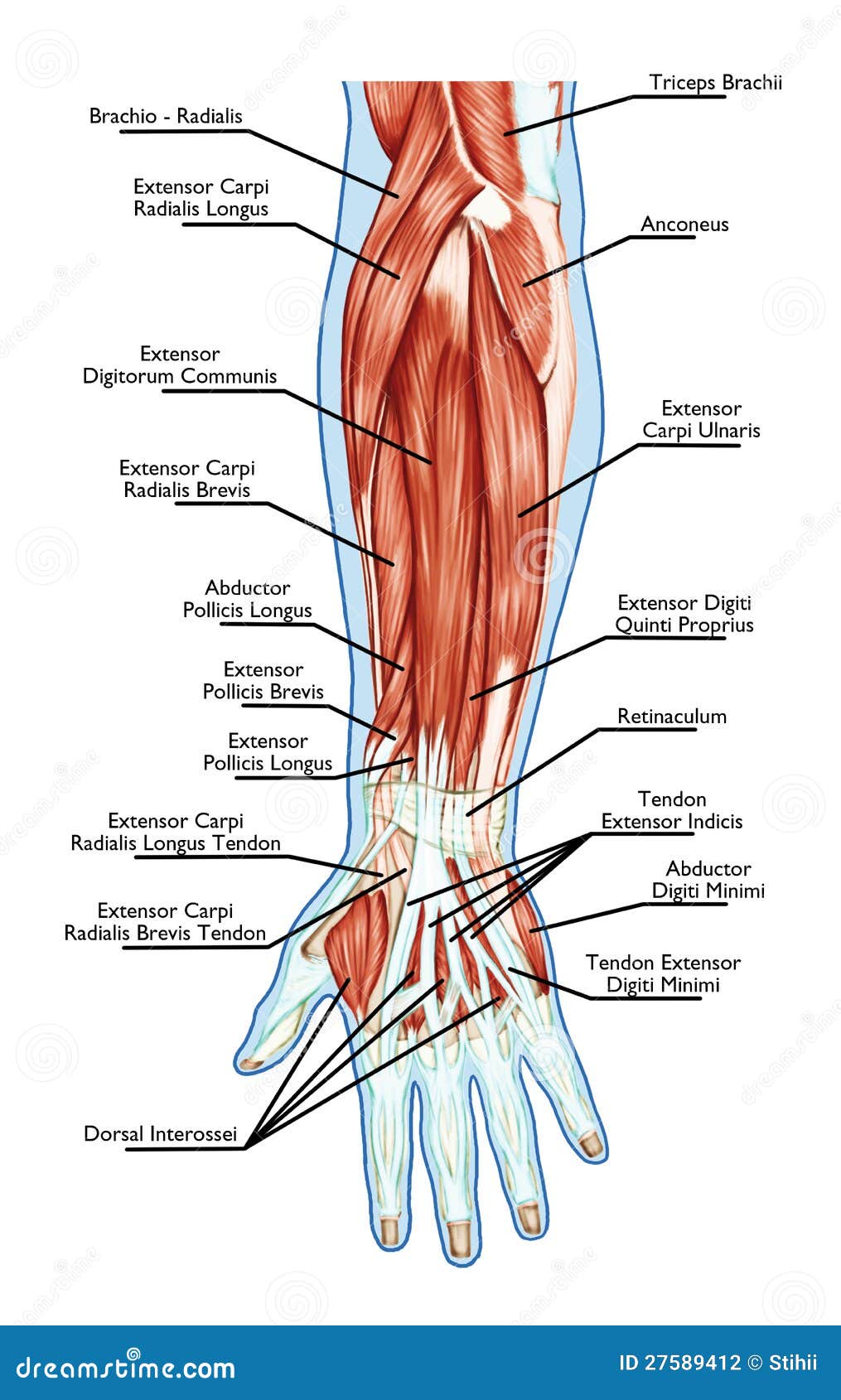
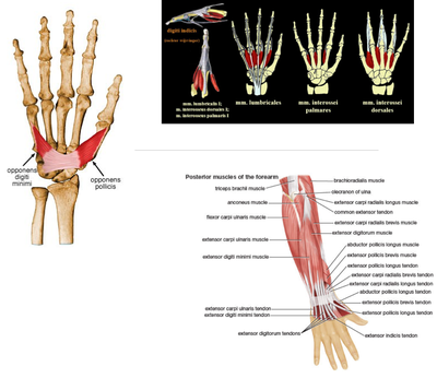
Together these combined tendons extend the fingers at the three finger joints. Its name means extensor of the digits which is why there were also extensor digitorum tendons in the feet. 51c from radial to ulnar they include the 1 abductor pollicis longus and extensor pollicis brevis 2 extensor carpi radialis longus and brevis 3 extensor pollicis longus 4 extensor digitorum and extensor indicis 5 extensor digiti minimi and 6 extensor carpi ulnaris. In the finger the ends of other tendons that start in the hand join with them to make the fingers move.
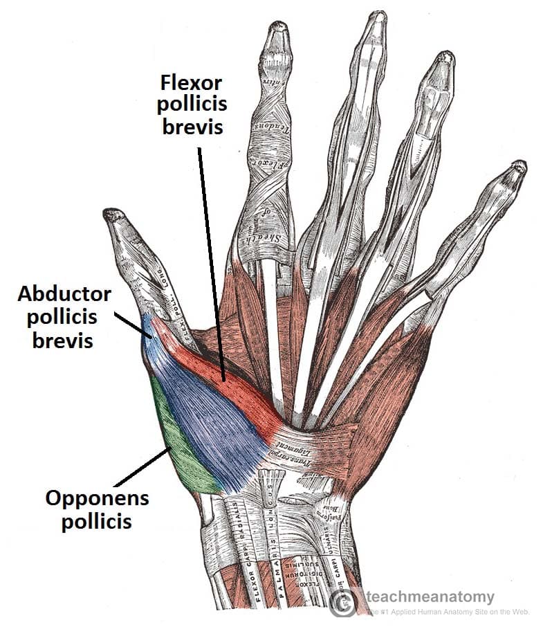
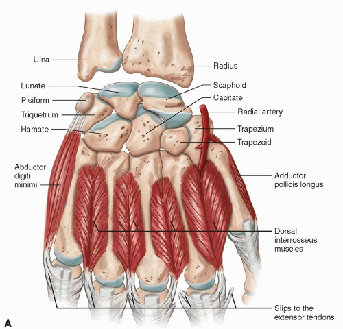
The extensor tendons are held in place by the extensor retinaculumas the tendons travel over the posterior back aspect of the wrist they are enclosed within synovial tendon sheaths. Extensor tendon compartments of the wrist are anatomical tunnels on the back of the wrist that contain tendons of muscles that extend as opposed to flex the wrist and the digits fingers and thumb. These muscles travel towards the hand where they eventually connect to the extensor tendons before crossing over the back of the wrist joint. We are pleased to provide you with the picture named hand muscle and tendon anatomy dorsal viewwe hope this picture hand muscle and tendon anatomy dorsal view can help you study and research.







:background_color(FFFFFF):format(jpeg)/images/library/10610/DorsalHand_Thumbnail.png)












:background_color(FFFFFF):format(jpeg)/images/article/en/dorsal-interossei-muscles-of-the-hand/waJ2TLmVQQXoNNOd22zgYA_Dorsal_interossei_muscles_of_the_hand.png)

